The Four Maps: What to Look at in Each
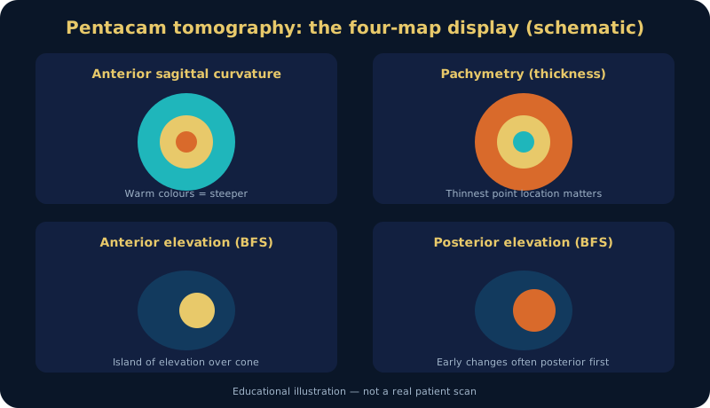
A standard Pentacam report shows four colour maps. Warm colours (red/orange) mean steeper curvature, thinner tissue or higher elevation depending on the map — colour alone never diagnoses.
- Anterior sagittal (axial) curvature: front-surface shape; asymmetric inferior steepening or a skewed bow-tie raises suspicion.
- Tangential curvature: more sensitive to sharp local change and shows the cone apex clearly.
- Pachymetry: thickness distribution and thinnest-point location; a displaced thinnest point with rapid peripheral progression matters.
- Elevation maps: height of the anterior and posterior surfaces above a reference shape — the most sensitive to early change.

Why the Back Surface Matters
Early posterior changes may precede obvious anterior change, so anterior maps are never read alone. Isolated borderline posterior elevation with a normal-looking front surface means suspicion and follow-up — not a final diagnosis or treatment decision.
Thickness Patterns Matter More Than One Number
There is no universal thickness cut-off for every eye; what matters is the pattern: where is the thinnest point? Is it displaced? Is the centre-to-periphery progression normal or steep? A constitutionally thin cornea can be entirely normal with regular maps and stability over time.
BAD-D and ARTmax: What They Add
- BAD (Belin/Ambrósio Enhanced Ectasia Display): combines several indices (anterior/posterior curvature, thickness distribution and more) into a composite reading; high values support suspicion but are interpreted within the full picture.
- ARTmax (Ambrósio Relational Thickness maximum): relates the thinnest pachymetry to the peripheral thickness gradient; very low values support an ectatic pattern.
These are powerful aids for screening and early diagnosis — not standalone verdicts.
Kmax Limits: Why It Is Not Enough Alone
- It measures maximum curvature at one point, affected by dry eye, tear instability and fixation during the scan.
- It may spike temporarily and return to normal next visit without any real disease change.
- Progression assessment needs consistent change in more than one parameter (posterior curvature, thickness, elevation) across serial scans on the same device.
Suspicion vs Diagnosis vs Progression
| Situation | What we see | Decision |
|---|---|---|
| Normal | Regular maps, indices within normal range, stability over time | Routine follow-up as appropriate |
| Suspect | An isolated sign (e.g. borderline posterior elevation) without the full pattern | Periodic follow-up, no pre-emptive treatment |
| Confirmed | Complete pattern: irregular curvature + displaced thinning + high elevation + clinical signs | Follow-up plan and cross-linking assessment |
| Documented progression | Consistent multi-parameter change between two reliable same-device scans | Discuss cross-linking — cross-linking guide |
Conditions for a Valid Comparison
- Same device, and no contact lenses for an adequate period before scanning.
- Good hydration and acceptable scan quality (stated in the report).
- Compare the maps themselves, not only summary numbers.
Brief Differential Diagnosis
Not every irregular curvature is keratoconus: post-surgical or post-injury astigmatism, pellucid marginal degeneration and long-term contact-lens warpage can mimic the pattern — hence rescanning after a lens holiday and correlating with clinical examination. See also: keratoconus library and biomechanics and epithelial mapping.
FAQs
Does a high Kmax mean I have keratoconus?
Not necessarily; it is one maximum-curvature point influenced by tears and dryness. Diagnosis needs the integrated picture with clinical examination.
Why does the doctor repeat the Pentacam after months?
Because proving progression requires reliable serial comparison — and that is what the cross-linking decision is built on.
Should I stop wearing lenses before the scan?
Yes, depending on lens type and wear history — ask the clinic when booking how long a lens holiday your case needs.
Does a red map mean advanced disease?
Red only means a higher value on the colour scale; severity is judged by the full pattern, thickness, vision and stability over time.
Selected references: Gomes JAP et al. Global consensus on keratoconus and ectatic diseases. Cornea. 2015;34(4):359–369. · Rabinowitz YS. Keratoconus. Surv Ophthalmol. 1998;42(4):297–319.
Related topics
Author: Prof. Dr. Amr Said — Assistant Professor of Ophthalmology, Faculty of Medicine, Alexandria University. Published November 2026. Last medical review: November 2026. Educational content; not a substitute for individual assessment.
Prof. Dr. Amr Said Cornea & Refractive Surgery Clinic
Prof. Dr. Amr Said — Assistant Professor of Ophthalmology, Faculty of Medicine, Alexandria University; consultant in corneal and refractive surgery, keratoconus treatment and cataract surgery.
Smouha branch: 5 Dr Sayed Fahmy Street, off Bahaa El-Din Ghatwary Street, Smouha — behind Sidi Gaber railway station. Branch map
Amreya branch: In front of Amreya bridge — Omar El-Mokhtar Tower, 3rd floor, Amreya. Branch map
Phones: 01226154154 · 01032037199 · 01021031651 · Amreya: 01096615751
Working hours and consultation fees are subject to updates — please confirm your appointment and fee by phone or WhatsApp before attending.