Cornea Centre Sections
Each section is a complete educational page linking back to this hub and to related steps in diagnosis and treatment.
On this page
Keratoconus Diagnosis: Pentacam & Tomography
Anterior/posterior curvature, pachymetry, elevation, BAD-D — and each number's limits.
Read diagnosis →Corneal Cross-Linking
Techniques, indications and treatment decisions: standard, accelerated, transepithelial.
Read cross-linking →Intracorneal Ring Segments
Indications, planning and expected outcomes — visual rehabilitation, not stabilisation.
Read rings →Specialty Contact Lenses
RGP, hybrid and scleral lenses: fitting, care and their role around surgery.
Read lenses →Advanced Keratoconus & Transplantation
When is transplantation needed? DALK versus penetrating keratoplasty.
Read transplantation →Paediatric Keratoconus
Early detection and management — with a parent FAQ.
Read paediatric →Biomechanics & Epithelial Mapping
Modern corneal diagnostics: biomechanical strength and epithelial thickness maps.
Read advanced →What Is Keratoconus?
Causes, early symptoms, diagnosis and the full treatment path.
Read overview →How to Read a Pentacam
The four maps, BAD-D, ARTmax, Kmax limits — and why one number never suffices.
Read the guide →Keratoconus in Children
Early detection and individualised treatment — a parent-friendly guide.
Read children's →Corneal Cross-Linking
Techniques, indications and decisions — in the English library.
Read cross-linking →Specialty Lenses & Glasses
RGP vs hybrid vs scleral — and when glasses suffice.
Read lenses →Transplantation: DALK vs PK
When is a graft needed? Layers, rejection, sutures, rehabilitation.
Read transplantation →Keratoconus & Pregnancy
Safe monitoring, deferring elective cross-linking, no LASIK in pregnancy.
Read pregnancy →iVIS Ray Tracing
Analysis and planning technology for keratoconus optics — role and limits.
Read ray tracing →Astigmatism
Regular vs irregular: symptoms, diagnosis and treatment options.
Read astigmatism →Keratoconus Library
Symptoms, causes, stages, eye rubbing, allergy and more.
Browse library →Why the Cornea Matters
The cornea is the eye's clear front window and provides roughly two-thirds of its refractive power — so shape, clarity and thickness changes directly affect vision.
Keratoconus and corneal ectasia
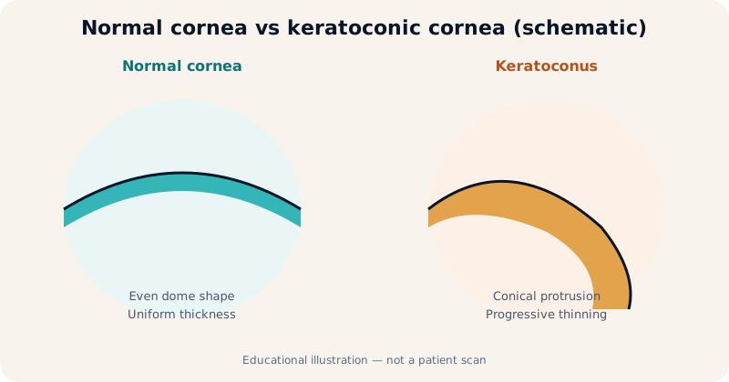
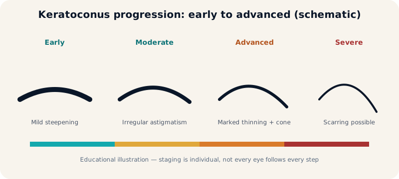
Keratoconus is a progressive thinning and conical protrusion, usually starting in the teens or twenties, causing irregular astigmatism that glasses correct poorly in advanced stages. Genetic and environmental factors contribute, notably chronic eye rubbing and allergy, with appropriate qualification of each factor's role.
Cornea and refractive surgery
A healthy cornea is a precondition for LASIK, Femto-SMILE and phakic lenses; tomography, biomechanics and epithelial mapping screen for hidden weakness that could lead to post-operative ectasia. See biomechanics and epithelial mapping.
Corneal Diagnostic Tests
Each test answers a different clinical question — diagnosis integrates them; no single number decides.
| Test | What it measures | Question it answers | Limitation |
|---|---|---|---|
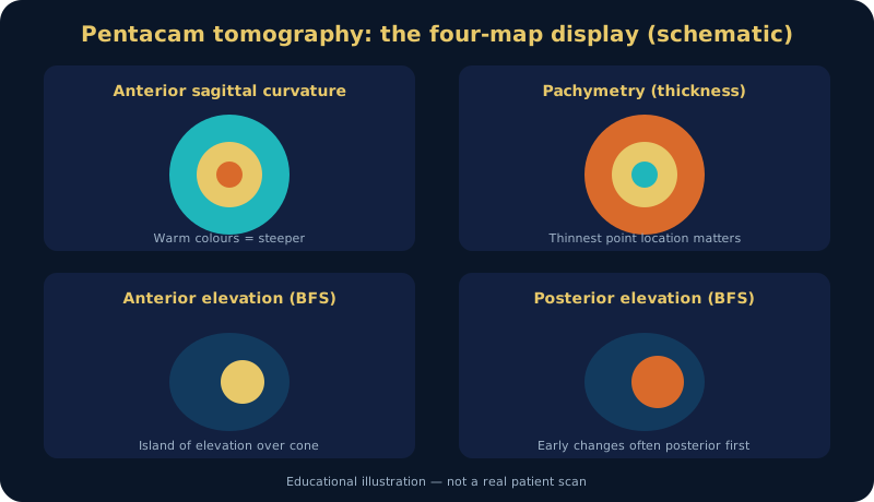
| Pentacam / tomography | Both surfaces, pachymetry, elevation | Is there keratoconus? Is it progressing? | Needs serial scans under similar conditions |
| Topography | Anterior surface only | Front-surface regularity | Blind to posterior surface and thickness |
| Biomechanics | Stiffness and deformation response | Hidden ectasia risk before LASIK | Never excludes risk alone |
| Epithelial mapping | Epithelial thickness distribution | Masked early ectasia | Always read with tomography |
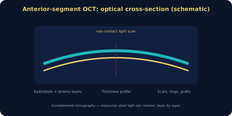
| Anterior-segment OCT | Optical layer cross-section | Scars, rings, grafts | Complement, not replacement |
Corneal Disease Directory
Beyond keratoconus — dedicated pages only where verified content exists.
Ulcers & Infections
Bacterial, fungal, viral and contact-lens ulcers: urgent diagnosis protects vision.
Read the directory →Penetrating Keratoplasty
Surgical principle, follow-up and graft rejection in detail.
Read transplantation →Dry Eye & Ocular Surface
A healthy surface underpins every corneal treatment and lens fit.
Read dry eye →Cataract with Corneal Disease
Lens and calculation decisions with keratoconus, scars or prior LASIK.
Read cataract-cornea →Research and Knowledge
Educational content connects where verified to the research of Prof. Dr. Amr Said (Assistant Professor of Ophthalmology, Faculty of Medicine, Alexandria University): paediatric keratoconus, cross-linking, biomechanics, tomography, rings and refractive safety. No study or result is attributed without verification.
When to Seek Corneal Assessment
- Repeated glasses changes or rising astigmatism, especially in teenagers.
- Blur despite new glasses, or new night halos and ghosting.
- Chronic eye rubbing with allergy.
- Family history of keratoconus, or LASIK plans with suspected irregularity.
- Lens intolerance or sudden deterioration in one eye (possible hydrops — urgent).
Clinic in Alexandria: Smouha and Amreya. International patients (Libya, Sudan, Gulf): see the international patients page — reports can be reviewed preliminarily, but never replace clinical examination.
Frequently Asked Questions
Direct answers — details in the linked pages.
How is keratoconus diagnosed?
Clinical examination plus Pentacam: both surfaces, pachymetry, elevation and indices such as BAD-D — with serial follow-up. Details (Arabic)
What is the difference between topography and tomography?
Topography maps the front surface only; tomography (Scheimpflug) measures both surfaces plus thickness — the standard for early ectasia. Details (Arabic)
When is corneal cross-linking recommended?
When progression is documented and safety criteria are met; decided per eye. Details (Arabic)
Can keratoconus be treated without transplantation?
Yes in most cases — cross-linking, lenses and rings; transplantation for advanced disease only. Details (Arabic)
Rings versus lenses — what is the difference?
Rings surgically regularise shape; lenses rehabilitate optics externally — neither halts progression. Details
Can children develop keratoconus?
Yes, sometimes faster; watch prescription changes and eye rubbing. Parent guide (Arabic)
How does biomechanics reveal ectasia risk?
It measures true corneal strength, not just thickness — read with tomography before LASIK. Details
What is epithelial mapping?
An epithelial thickness map showing masking around the cone (thinning over the apex). Details
When is corneal transplantation required?
With severe scarring or failed lens rehabilitation. Details (Arabic)
Selected References
- Rabinowitz YS. Keratoconus. Survey of Ophthalmology. 1998;43(2):121-140.
- Wollensak G, Spoerl E, Seiler T. Riboflavin/ultraviolet-A-induced collagen crosslinking for the treatment of keratoconus. Am J Ophthalmol. 2003;135(5):620-627.
- Belin MW, Ambrósio R. Scheimpflug imaging for keratoconus and ectatic disease. Indian J Ophthalmol. 2013;61(8):401-406.
- Global consensus on keratoconus and ectatic diseases (Gomes et al.). Cornea. 2015;34(4):359-369.
- Ferdi AC, et al. Natural history of keratoconus. Cornea. 2019 — plus recent systematic reviews on cross-linking and rings.
Author: Prof. Dr. Amr Said — Assistant Professor of Ophthalmology, Faculty of Medicine, Alexandria University. Published November 2026. Educational content; not a substitute for clinical examination.
Book a Cornea Assessment in Alexandria
Pentacam examination, cross-linking consultation or specialty-lens assessment — Smouha and Amreya.
Prof. Dr. Amr Said Cornea & Refractive Surgery Clinic
Prof. Dr. Amr Said — Assistant Professor of Ophthalmology, Faculty of Medicine, Alexandria University; consultant in corneal and refractive surgery, keratoconus treatment and cataract surgery.
Smouha branch: 5 Dr Sayed Fahmy Street, off Bahaa El-Din Ghatwary Street, Smouha — behind Sidi Gaber railway station. Branch map
Amreya branch: In front of Amreya bridge — Omar El-Mokhtar Tower, 3rd floor, Amreya. Branch map
Phones: 01226154154 · 01032037199 · 01021031651 · Amreya: 01096615751
WhatsApp consultation Call: 01226154154 Patient reviews
Working hours and consultation fees are subject to updates — please confirm your appointment and fee by phone or WhatsApp before attending.