
Advanced Disease: When to Consider a Graft
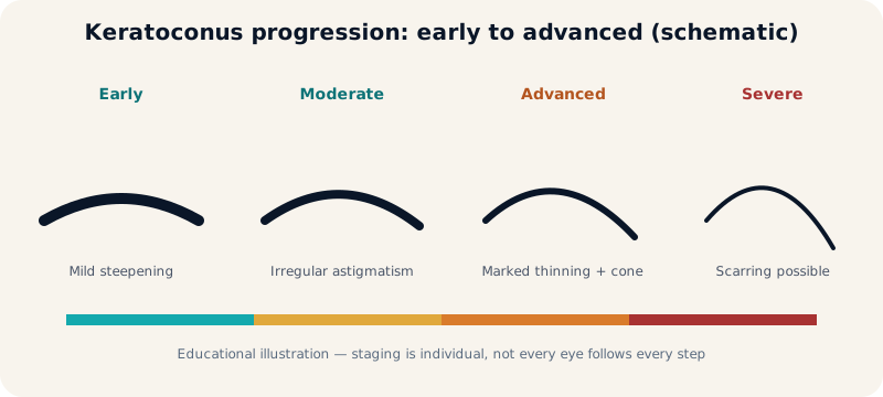
- Severe irregularity with marked thinning and poor vision despite specialty lenses.
- Central scarring blocking the visual axis.
- Contact-lens intolerance with no other rehabilitative option.
- Acute hydrops: managed first; transplantation discussed after stabilisation where indicated.
Transplantation is not automatic in advanced keratoconus — many advanced eyes are still rehabilitated with scleral lenses.
DALK vs Penetrating Keratoplasty
| Aspect | DALK (deep anterior lamellar) | PK (penetrating) |
|---|---|---|
| Layers replaced | Front stroma; patient's own Descemet + endothelium kept | Full thickness including donor endothelium |
| Endothelial rejection | Generally lower (own endothelium kept) | Higher; lifelong endothelial vigilance |
| Typical use | Healthy endothelium, no deep scarring | Deep scarring, endothelial disease, failed prior graft assessment |
| Wound / sutures | Structurally stronger eye; sutures still needed | Full-thickness wound; longer suture dependence |
Rejection and Graft Failure
- Rejection can affect the epithelium, stroma or (in PK) endothelium; DALK reduces but does not eliminate the risk.
- Warning signs — redness, pain, light sensitivity, vision drop: urgent review, never self-treatment with old drops.
- Failure (irreversible clouding) is managed per cause: medications, repeat graft assessment, or lens rehabilitation.
After Surgery: Sutures and Visual Rehabilitation
- Sutures stay months to over a year, adjusted or removed selectively for astigmatism.
- Vision is rehabilitated gradually: glasses, then specialty lenses for residual astigmatism once healed.
- Lifelong periodic follow-up: pressure, endothelium (PK), surface and suture checks.
FAQs
Will I see perfectly after transplantation?
No specific outcome is promised; most grafts restore useful vision with correction, and rehabilitation continues after healing.
How long does a graft last?
Many grafts serve for decades with care; longevity depends on technique, rejection vigilance and follow-up — no graft survival is guaranteed.
Can keratoconus return in the graft?
Recurrence in donor tissue is uncommon and late; lifelong follow-up detects change early.
Are lenses needed after a graft?
Often yes for residual astigmatism once healed — fitted individually.
Selected references: Watson SL, Ramsay A, Dart JKG, Bunce C, Craig E. Comparison of deep lamellar keratoplasty and penetrating keratoplasty in patients with keratoconus. Ophthalmology. 2004;111(9):1676–1682. · Sugita J, Kondo J. Deep lamellar keratoplasty with complete removal of pathological stroma for vision improvement. Br J Ophthalmol. 1997;81(3):184–188.
Related topics
Author: Prof. Dr. Amr Said — Assistant Professor of Ophthalmology, Faculty of Medicine, Alexandria University. Published November 2026. Last medical review: November 2026. Educational content; not a substitute for individual assessment.
Prof. Dr. Amr Said Cornea & Refractive Surgery Clinic
Prof. Dr. Amr Said — Assistant Professor of Ophthalmology, Faculty of Medicine, Alexandria University; consultant in corneal and refractive surgery, keratoconus treatment and cataract surgery.
Smouha branch: 5 Dr Sayed Fahmy Street, off Bahaa El-Din Ghatwary Street, Smouha — behind Sidi Gaber railway station. Branch map
Amreya branch: In front of Amreya bridge — Omar El-Mokhtar Tower, 3rd floor, Amreya. Branch map
Phones: 01226154154 · 01032037199 · 01021031651 · Amreya: 01096615751
Working hours and consultation fees are subject to updates — please confirm your appointment and fee by phone or WhatsApp before attending.