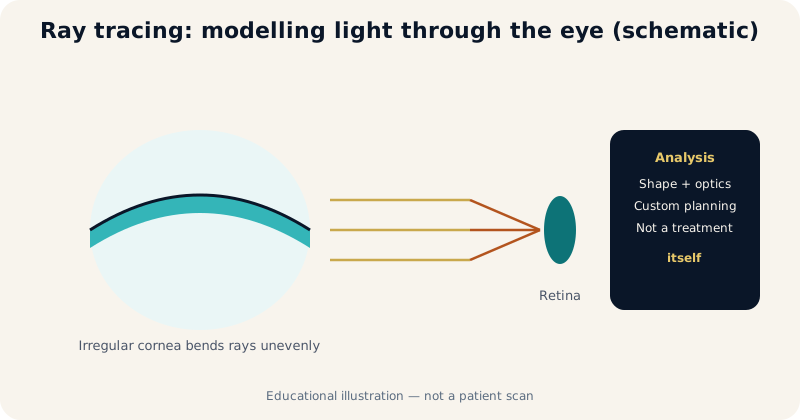
What Happens in Keratoconus Optics
The weakened cornea bulges irregularly, producing irregular astigmatism and distorted ray paths. Hence the classic complaint: "I see with glasses, but the picture is never sharp" — with ghosting and halos, especially in dim light.
Where Ray Tracing Fits
Instead of asking only "how many dioptres?", ray tracing asks: what happens to light entering this eye, and how do corneal shape and aberrations affect the retinal image? That matters because the link between corneal shape and image quality is complex in irregular corneas.
What It Does Not Do
- It does not stop keratoconus — cross-linking is the stabilising procedure where progression is proven.
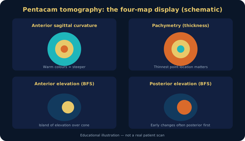
- It does not replace examination, Pentacam, refraction, surface assessment or history.
- Platform and device availability (including the iRES excimer platform) are discussed in clinic according to what is installed and approved — no device claim is made here.
Why It Matters After Cross-Linking
After stabilisation, irregular astigmatism and aberrations often remain: the disease stopped, but quality did not normalise. Advanced optical analysis then helps plan the second stage — visual rehabilitation with lenses or rings in selected eyes.
Refractive Surgery and Complex Cases
Keratoconus is never handled as "−4 dioptres, correct −4". In selected stable eyes after full assessment, advanced optical data may help design customised correction — but decisions rest on the complete dataset (Pentacam, examination, refraction, surface, acuity, history), never on ray tracing alone. In complex cases needing specialty lenses, understanding the optical system also guides expectations.
FAQs
Is ray tracing a laser treatment?
No — it is measurement and modelling. Any laser step is a separate procedure with its own indications, discussed only after full assessment.
Does every patient need it?
No — selected cases where full-system optics change planning.
What should I bring to assessment?
Old scans and prescriptions (they document history), a list of drops, and lenses removed for the advised period.
Selected references: Gomes JAP et al. Global consensus on keratoconus and ectatic diseases. Cornea. 2015;34(4):359–369. · Rabinowitz YS. Keratoconus. Surv Ophthalmol. 1998;42(4):297–319.
Related topics
Author: Prof. Dr. Amr Said — Assistant Professor of Ophthalmology, Faculty of Medicine, Alexandria University. Published November 2026. Last medical review: November 2026. Educational content; not a substitute for individual assessment.
Prof. Dr. Amr Said Cornea & Refractive Surgery Clinic
Prof. Dr. Amr Said — Assistant Professor of Ophthalmology, Faculty of Medicine, Alexandria University; consultant in corneal and refractive surgery, keratoconus treatment and cataract surgery.
Smouha branch: 5 Dr Sayed Fahmy Street, off Bahaa El-Din Ghatwary Street, Smouha — behind Sidi Gaber railway station. Branch map
Amreya branch: In front of Amreya bridge — Omar El-Mokhtar Tower, 3rd floor, Amreya. Branch map
Phones: 01226154154 · 01032037199 · 01021031651 · Amreya: 01096615751
Working hours and consultation fees are subject to updates — please confirm your appointment and fee by phone or WhatsApp before attending.